
Mouse Secondary Antibodies


Biotium Ferritin, Light Chain (FTL) (Microglia Marker) (FTL/1386), CF568 conjugate, 0.1mg/mL
Mammalian ferritins consist of 24 subunits made up of 2 types of polypeptide chains, ferritin heavy chain and ferritin light chain. Ferritin heavy chains catalyze the first step in iron storage, the oxidation of Fe (II), whereas ferritin light chains promote the nucleation of ferrihydrite, enabling storage of Fe (III). Light chain ferritin is involved in cataracts by at least two mechanisms, hereditary hyperferritinemia cataract syndrome, in which light chain ferritin is overexpressed, and oxidative stress, an important factor in the development of ageing-related cataracts.Primary antibodies are available purified, or with a selection of fluorescent CF Dyes and other labels. CF Dyes offer exceptional brightness and photostability. Note: Conjugates of blue fluorescent dyes like CF405S and CF405M are not recommended for detecting low abundance targets, because blue dyes have lower fluorescence and can give higher non-specific background than other dye colors.

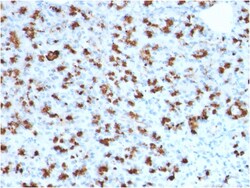
Biotium BARX1 (Prognostic Biomarker in Hepatocellular Carcinoma)(BARX1/2760), 1mg/mL
BarX1 is a member of the Bar subclass of homeobox transcription factors. Studies of the Mouse and chick homolog suggest the encoded protein may play a role in developing teeth and craniofacial mesenchyme of neural crest origin. The protein may also be associated with differentiation of stomach epithelia. Down-regulation of Barx1 promotes hepatocellular carcinoma cell (HCC) migration, invasion and metastasis, whereas up-regulation of Barx1 inhibits HCC migration, invasion and metastasis. The loss of Barx1 expression represents a prognostic biomarker in human HCC. Primary antibodies are available purified, or with a selection of fluorescent CF Dyes and other labels. CF Dyes offer exceptional brightness and photostability. Note: Conjugates of blue fluorescent dyes like CF405S and CF405M are not recommended for detecting low abundance targets, because blue dyes have lower fluorescence and can give higher non-specific background than other dye colors.
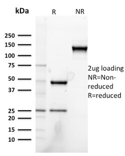
Biotium Heregulin-1 / Neuregulin-1 (Breast and Urothelial Marker)(NRG1/2752), Biotin conjugate, 0.1mg/mL
Heregulin-1 is a membrane glycoprotein that mediates cell-cell signaling and plays a critical role in the growth and development of multiple organ systems. An extraordinary variety of different isoforms are produced from this gene through alternative promoter usage and splicing. These isoforms are expressed in a tissue-specific manner and differ significantly in their structure, and are classified as types I, II, III, IV, V and VI. Dysregulation of this gene has been linked to diseases such as cancer, schizophrenia, and bipolar disorder (BPD). Primary antibodies are available purified, or with a selection of fluorescent CF Dyes and other labels. CF Dyes offer exceptional brightness and photostability. Note: Conjugates of blue fluorescent dyes like CF405S and CF405M are not recommended for detecting low abundance targets, because blue dyes have lower fluorescence and can give higher non-specific background than other dye colors.
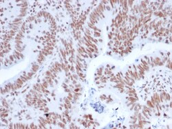
Biotium NRF1(NRF1/2608), CF568 conjugate, 0.1mg/mL
The NF-E2 DNA binding protein is composed of two subunits, p45 and MafK, and it regulates expression of globin genes in developing erythroid cells through interaction with Maf recognition elements (MAREs). A family of NF-E2 related proteins, which are collectively known as the Cap 'n' collar (CNC) family and include Nrf1 (also designated TCF11), Nrf2 and Nrf3, are bZIP transcription factors that heterodimerize with Maf proteins to bind MARE sequences. The Nrf proteins also bind the antioxidant response element (ARE) and are implicated in the regulation of detoxification enzymes and the oxidative stress response. They do so by heterodimerizing with Jun family members (c-Jun, JunB and JunD) to activate gene expression, specifically the detoxifying enzyme, NQO1. Nrf2 is widely expressed and is thought to translocate to the nucleus after treatment with xenobiotics and antioxidants, which stimulate its release from a repressor protein Keap1. Nrf3 is highly expressed in placenta, B
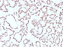
Biotium Cyclin D1 (G1-Cyclin & Mantle Cell Lymphoma Marker)(CCND1/2593), CF568 conjugate, 0.1mg/mL
Recognizes a protein of 36 kDa, identified as cyclin D1. Cyclin D1, one of the key cell cycle regulators, is a putative proto-oncogene overexpressed in a wide variety of human neoplasms. This antibody neutralizes the activity of cyclin D1 in vivo. About 60% of mantle cell lymphomas (MCL) contain a t(11; 14)(q13; q32) translocation resulting in over-expression of cyclin D1. This antibody is useful in identifying mantle cell lymphomas (cyclin D1 positive) from CLL/SLL and follicular lymphomas (cyclin D1 negative). Occasionally, hairy cell leukemia and plasma cell myeloma weakly express Cyclin D1. Primary antibodies are available purified, or with a selection of fluorescent CF Dyes and other labels. CF Dyes offer exceptional brightness and photostability. Note: Conjugates of blue fluorescent dyes like CF405S and CF405M are not recommended for detecting low abundance targets, because blue dyes have lower fluorescence and can give higher non-specific background than other dye
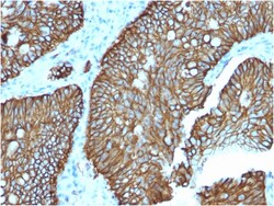
Biotium Cadherin 17 / LI Cadherin (Liver-Intestine Marker)(CDH17/2618), CF488A conjugate, 0.1mg/mL
Cadherin 17, also known as LI cadherin (for liver-intestine cadherin), is a member of the cadherin family of calcium-dependent adhesion molecules. Cadherins mediate cell-cell interactions via hemophilic binding, and are critical to the maintenance of tissue structure and morphogenesis. Cadherins each contain a large extracellular domain at the amino terminus, which is characterized by a series of five homologous repeats, the most distal of which is thought to be responsible for binding specificity. The relatively short carboxy terminal, intracellular domain interacts with a variety of cytoplasmic proteins, including beta-catenin, to regulate cadherin function. Cadherin 17 expression is restricted to liver and intestine tissues and is specifically localized to the basolateral domain of hepatocytes and enterocytes. Primary antibodies are available purified, or with a selection of fluorescent CF Dyes and other labels. CF Dyes offer exceptional brightness and photostability. N
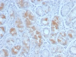
Biotium Gastrin(GAST/2634), CF488A conjugate, 0.1mg/mL
Gastrin, is a hormone that normally formed by mucosal cells in the gastric antrum and by the D cells of the pancreatic islets. Its primary function is to stimulate secretion of HCl by the gastric mucosa. HCl, in turn, inhibits gastrin formation. It also responsible for stimulating smooth muscle contraction and increasing blood circulation and water secretion in the stomach and intestine. Gastrin is regulated by epidermal growth factor in both mice and humans. Gastrin is excreted in excess by pancreatic tumors in the Zollinger-Ellison syndrome. Gastrin-Releasing Peptide (GRP) stimulates the release of gastrin as well as other gastrointestinal hormones and also acts as an autocrine growth factor for certain cell types. High levels of GRP are found in the human lung just after birth and levels decrease thereafter in parallel with the observed disease in a number of pulmonary neuroendocrine cells. GRP is known to promote lung tumorigenesis in model systems. Primary antibodies are
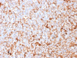
Biotium Mammaglobin (SCGB2A2) (Breast Cancer Marker)(MGB1/2682R), CF488A conjugate, 0.1mg/mL
Mammaglobin is a 93 amino acid glycoprotein with homology to other secretoglobin-uteroglobin family members. It was originally identified as a breast cancer restricted biomarker by differential screening. Mammaglobin related to secretoglobin family that includes human uteroglobin and lipophilin. Mammaglobin antibody stains cytoplasm of normal breast epithelial cells as well as primary and metastatic breast carcinomas. Mammaglobin expression is absent in prostate, kidney, colon, rectum, small intestine, stomach, pancreas, lung, and thyroid tissues. Mammaglobin may be used as part of an immunohistochemical panel for determination of metastatic breast carcinoma and tumor of unknown primary origin. Primary antibodies are available purified, or with a selection of fluorescent CF Dyes and other labels. CF Dyes offer exceptional brightness and photostability. Note: Conjugates of blue fluorescent dyes like CF405S and CF405M are not recommended for detecting low abundance targets
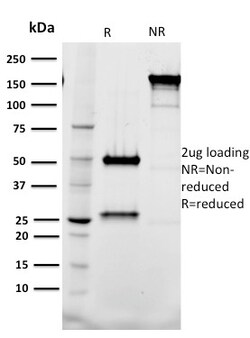
Biotium Tal1(BTL73), 0.2mg/mL
Activation of TAL1 characterizes up to 60% of cases of human T cell acute lymphoblastic leukemia, making it the most frequent gain-of-function mutation observed in this disorder. TAL1 (also designated SCL) is a serine phosphoprotein and basic helix-loop-helix transcription factor known to regulate embryonic hematopoiesis. This transcription factor binds as a heterodimer with E2A and HEB/HTF4 to a nucleotide sequence motif termed the E-box. In addition, leukemogenesis is accelerated dramatically by transgenic co-expression of TAL1 and the catalytic subunit of casein kinase IIHLH transcription factors. Primary antibodies are available purified, or with a selection of fluorescent CF Dyes and other labels. CF Dyes offer exceptional brightness and photostability. Note: Conjugates of blue fluorescent dyes like CF405S and CF405M are not recommended for detecting low abundance targets, because blue dyes have lower fluorescence and can give higher non-specific background than oth
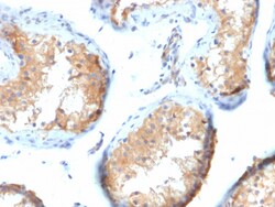
Biotium Vinculin (Marker of Age-related Macular Degeneration)(VCL/2573), 1mg/mL
Focal adhesions are identified as areas within the plasma membrane of tissue culture cells that adhere tightly to the underlying substrate. In vivo, these regions are involved in the adhesion of cells to the extracellular matrix. Paxillin and vinculin are cytoskeletal, focal adhesion proteins that are components of a protein complex which links the Actin network to the plasma membrane. Vinculin binding sites have been identified on other cytoskeletal proteins, including talin and alpha-actinin each contain actin binding sites. Reportedly, vinculin is a potential plasma biomarker for Age-related Macular Degeneration (AMD). The early detection of AMD using novel plasma biomarkers with genetic modeling may enable timely treatment and vision preservation in the elderly. Primary antibodies are available purified, or with a selection of fluorescent CF Dyes and other labels. CF Dyes offer exceptional brightness and photostability. Note: Conjugates of blue fluorescent dyes like CF
Biotium Ki-67 (Proliferating Cell Marker) (MKI67/2465), Biotin conjugate, 0.1mg/mL
Ki-67 antigen is a nuclear, non-histone protein that is present in all stages of the cell cycle except G0. This characteristic makes Ki-67 an excellent marker for proliferating cells and is commonly used as one of the prognostic factors in cancer studies. A correlation has been demonstrated between Ki-67 index and the histo-pathological grade of neoplasms. Assessment of Ki-67 expression in renal and ureter tumors shows a correlation between tumor proliferation and disease progression, thus making it possible to differentiate high-risk patients. Ki-67 expression may also prove to be important for distinguishing between malignant and benign peripheral nerve sheath tumors. Ki-67 labeling index has been shown to be a prognostic marker in a number of neoplasms including grade II astrocytoma, oligodendroglioma, colon carcinoma, and breast carcinoma. In general, Ki-67 is a good marker of proliferating cell populations.Primary antibodies are available purified, or with a selection of
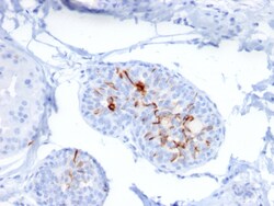
Biotium ZNF690 / ZSCAN29(ZSCAN29/2610), Biotin conjugate, 0.1mg/mL
Zinc finger proteins contain DNA-binding domains and have a wide variety of functions, most of which encompass some form of transcriptional activation or repression. The majority of zinc finger proteins contain a Kruppel-type DNA binding domain and a KRAB domain, which is thought to interact with KAP1, thereby recruiting histone modifying proteins. Zinc finger protein 690 (ZNF690), also known as ZSCAN29, is a 851 amino acid member of the Kruppel C2H2- type zinc finger protein family. Localized to the nucleus, ZNF690 contains six C2H2-type zinc fingers and one KRAB domain through which it is thought to be involved in DNA-binding and transcriptional regulation. Four isoforms of ZNF690 exist as a result of alternative splicing events. Primary antibodies are available purified, or with a selection of fluorescent CF Dyes and other labels. CF Dyes offer exceptional brightness and photostability. Note: Conjugates of blue fluorescent dyes like CF405S and CF405M are not recommend
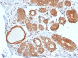
Biotium STAT5B(STAT5B/2611), CF488A conjugate, 0.1mg/mL
Signal transducer and activator of transcription 5A (Stat5a) and Stat5b, which share 96% homology, undergo receptor tyrosine kinase or G protein-coupled receptor-dependent phosphorylation in response to cytokines or growth factors, and then form homo- or heterodimers that translocate to the nucleus, where they initiate transcription. Activation of Stat5a via IL-2, IL-3, IL-7/ GM-CSF, erythropoietin, thrombopoietin and growth hormones influences proliferation, differentiation and apoptosis in lymphohematopoietic cells. Phosphorylation of Stat5a at Ser127/Ser128 and Ser779 are contigent on ErbB-4-mediated activation of Stat5a. Activation of Stat5b via IL-2, IL-4, CSF-1 and growth hormones influences TCR signaling, apoptosis, adult mammary gland development and sexual dimorphism of liver gene expression. Stat5b is the major liver-expressed Stat5 form that has been shown to fuse with the retinoic acid receptor a gene in acute promyelocytic leukemias (APLL). Stat5a/b null mice hav
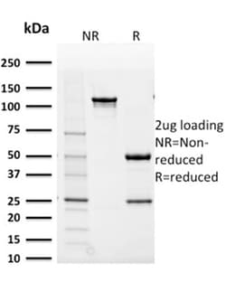
Biotium Aldo-keto Reductase Family 1 Member B1 (Adrenal Marker)(CPTC-AKR1B1-2), Biotin conjugate, 0.1mg/mL
Aldo-keto Reductase Family 1 Member B1 (AKR1B1), also known as aldose reductase or aldehyde reductase, is a member of the aldo/keto reductase superfamily, which consists of more than 40 known enzymes and proteins. This protein catalyzes the reduction of a number of aldehydes, including the aldehyde form of glucose, and is thereby implicated in the development of diabetic complications by catalyzing the reduction of glucose to sorbitol. It has also been shown to have decreased expression in adrenocortical cancer, and possibly play a role in adrenal tumorigenesis. It has been suggested that AKR1B1 could be investigated as a marker of malignancy for adrenal tumor diagnosis. Primary antibodies are available purified, or with a selection of fluorescent CF Dyes and other labels. CF Dyes offer exceptional brightness and photostability. Note: Conjugates of blue fluorescent dyes like CF405S and CF405M are not recommended for detecting low abundance targets, because blue dyes have
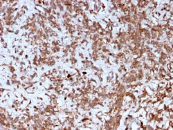
Biotium Cathepsin K (Marker of Tumor Invasiveness)(CTSK/2792), CF568 conjugate, 0.1mg/mL
Cathepsin K is a lysosomal and secreted cysteine proteinase involved in bone remodeling and resorption. This protein, which is a member of the peptidase C1 protein family, is predominantly expressed in osteoclasts. However, the encoded protein is also expressed in a significant fraction of human breast cancers, where it could contribute to tumor invasiveness. Mutations in this gene are the cause of pycnodysostosis, an autosomal recessive disease characterized by osteosclerosis and short stature. Primary antibodies are available purified, or with a selection of fluorescent CF Dyes and other labels. CF Dyes offer exceptional brightness and photostability. Note: Conjugates of blue fluorescent dyes like CF405S and CF405M are not recommended for detecting low abundance targets, because blue dyes have lower fluorescence and can give higher non-specific background than other dye colors.